
|
|
Wei Lu,
Bioengineering Grad. Student
Bryan Cunitz, Engineer
Peter Kaczkowski, Engineer
Oleg Sapozhnikov, Engineer
Mike Bailey, Engineer
Center for Industrial &
Medical Ultrasound
Applied Physics Laboratory
University of Washington
Anup Shah
Resident, Dept. of Urology
UW School of Medicine
NIH
NSBRI
|
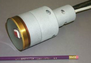
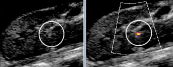
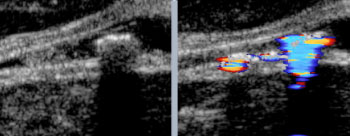
We built a new system that combines a commercial ultrasound imager for visualizing the stone and a focused ultrasound device for pushing the stone. The active end is a hand held applicator that is put in contact with the patient's skin and manipulated to find and then push the stone. Two conventional and readily available imaging modes are used: brightness or B-mode, and Color Flow Doppler mode. Brightness shows the bright reflection from the stones, which are hard compared to the surrounding tissue and fluid. In Doppler, the stone appears as a bright spot of rapidly changing color , and can be targeted using this "twinkling artifact." Doppler is usually used to detect blood flow and then label it with red or blue color on the image. With twinkling artifact, something in the ultrasound reflection from the stone confuses the Doppler processing and causes the imager to display the stone as a flickering mosaic of color. Natural and artificial stones were implanted in tissue-mimicking phantoms and in porcine models, targeted with ultrasound guidance, pushed with focused ultrasound radiation force, and the stone movement recorded with x-ray fluoroscopy and ultrasound. Stones were commonly moved 1-2 centimeters in about 1 second and were moved from the lower pole to the kidney exit, the ureteropelvic junction, or even into the ureter, the tube leading to the bladder. |
   |
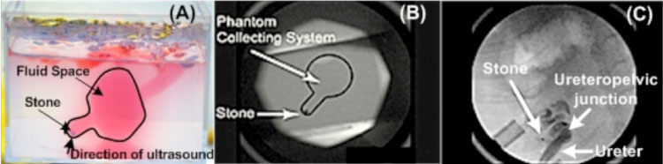 Above: A)The phantom is a translucent gel with red-colored water in a space mimicking the lower and mid poles of the kidney. The 3-mm stone rests in the bottom of the small chamber and is pushed up into the larger chamber. A) and B) The lower pole of the kidney and the stone are down and to the left in the images and the ultrasound pushes the stone up to the right, and in panel (C) actually out of the kidney into the ureter. |