|
|
Ultrasonic Detection, Propulsion + Comminution of Kidney Stones
Current Research at the University of Washington
|
|
We report results from several clinical trials of our investigational device that is an alternative to surgery for fragmenting and facilitating clearance of small, asymptomatic renal stones. Our office-based unltrasound system images, breaks, and repositions stone fragments to facilitate their natural clearance in the urine.
We have now compiled a data base of over 200 study participants who have undergone a procedure with these investigational technologies without serious adverse events.
|
|
Ultrasonic Propulsion Clears Residual Stone Fragments
|
|
|
|
|
|
A study conducted at the University of Washington and the VA of Puget Sound demonstrates that ultrasonic propulsion of residual kidney stone fragments reduces relapse for patients who have undergone stone intervention treatments.
The investigational team reports results of a trial conducted with 82 people from 2015 to 2024. Repositioning residual fragments in the treatment group results in a 70% lower incidence of relapse — urgent medical visit or a subsequent surgery. Time to relapse was also longer by nearly 1.5 years in the treatment group.
"This study is the culmination of our work to invent ultrasonic propulsion to remove kidney stone fragments. Our treatment technology and clinical methods reduce the number of patients who return to the emergency room or their urologist with stone problems." — Mike Bailey
|
|
|
 |
|
Randomized controlled trial of ultrasonic propulsion-facilitated clearance of residual kidney stone fragments vs. observation Sorensen, M.D., and 16 others including B. Dunmire, J. Thiel, B.W. Cunitz, J.C. Kucewicz, and M.R. Bailey, "Randomized controlled trial of ultrasonic propulsion-facilitated clearance of residual kidney stone fragments vs. observation," J. Urol., 212, 811-820, doi:10.1097/JU.0000000000004186, 2024. |
|
More Info
| |
|
1 Dec 2024 
|
|
 |
|
|
Ultrasonic propulsion is an investigational procedure for awake patients. Our purpose was to evaluate whether ultrasonic propulsion to facilitate residual kidney stone fragment clearance reduced relapse.
This multicenter, prospective, open-label, randomized, controlled trial used single block randomization (1:1) without masking. Adults with residual fragments (individually 5 mm or smaller) were enrolled. Primary outcome was relapse as measured by stone growth, a stone-related urgent medical visit, or surgery by 5 years or study end. Secondary outcomes were fragment passage within 3 weeks and adverse events within 90 days. Cumulative incidence of relapse was estimated using the Kaplan-Meier method. Log-rank test was used to compare the treatment (ultrasonic propulsion) and control (observation) groups.
The trial was conducted from May 9, 2015, through April 6, 2024. Median follow-up (interquartile range) was 3.0 (1.8–3.2) years. The treatment group (n = 40) had longer time to relapse than the control group (n = 42; P < .003). The restricted mean time-to-relapse was 52% longer in the treatment group than in the control group (1530 ± 92 days vs 1009 ± 118 days), and the risk of relapse was lower (hazard ratio 0.30, 95% CI 0.13–0.68) with 8 of 40 and 21 of 42 participants, respectively, experiencing relapse. Omitting 3 participants not asked about passage, 24 treatment (63%) and 2 control (5%) participants passed fragments within 3 weeks of treatment. Adverse events were mild, transient, and self-resolving, and were reported in 25 treated participants (63%) and 17 controls (40%).
|
|
|
|
Human Study Combines BWL and UP Treatment
|
|
|
|
|
|
The Laboratory and University research team demonstrates that burst wave lithotripsy (BWL) followed by ultrasonic propulsion (UP) in a clinical setting clears over 70% of asymtomatic, small renal stones in awake patients. This is the first human feasibility study that uses both technologies together to clear stones.
As in preceding trials, all participants tolerated the procedure without anesthesia or medication.
Based on this successful study, the team anticipates a larger efficacy trial.
|
|
|
 |
|
Facilitated clearance of small, asymptomatic renal stones with burst wave lithotripsy and ultrasonic propulsion Harper, J.D., and 18 others including B. Dunmire, J. Thiel, Y.-N. Wang, S. Totten, J.C. Kucewicz, and M.R. Bailey, "Facilitated clearance of small, asymptomatic renal stones with burst wave lithotripsy and ultrasonic propulsion," J. Urol., 214, 41-47, doi:10.1097/JU.0000000000004533, 2025. |
|
More Info
| |
|
1 Jul 2025 
|
|
 |
|
|
We tested feasibility of burst wave lithotripsy (BWL) and ultrasonic propulsion to noninvasively fragment and expel small, asymptomatic renal stones in awake participants.
Adult patients suspected of having 2- to 7-mm stones were consented and screened for eligibility. BWL and ultrasonic propulsion were applied to up to 3 stones in 1 kidney of qualifying participants for a 30-minute total exposure. Participants completed a CT scan and the Wisconsin Stone Quality-of-Life (WISQOL) questionnaire within 90 days before and 120 days after the procedure. Participants were contacted weekly for 3 weeks after the procedure to assess adverse events (AEs). Outcomes included (1) no fragment > 2 mm, (2) unanticipated health care visits, (3) change in stone volume, (4) reported AEs, and (5) WISQOL score.
Forty-one participants were enrolled between April 2023 and October 2024. Twenty-one participants failed screening because no stones were seen, stones were too large or small, stone visibility was too deep or obstructed, or they declined to participate. Twenty participants with 31 stones received the research procedure with 7 undergoing a single repeat procedure. Twenty-two of 31 stones (71%) met the primary effectiveness outcome of no fragment > 2 mm, with 17 of 31 stones (55%) reported as stone free. Median stone volume reduction (IQR) was 100% (88%–100%). No participants returned unexpectedly for care related to the procedure. AEs were all Grade I by modified Clavien classification. WISQOL scores improved on 10 of 15 completed questionnaires.
Small, asymptomatic renal stones were effectively and safely removed in awake participants in a clinic setting.
|
|
|
|
Indpendent Replication of Results
|
|
|
|
A randomized controlled trial of ultrasonsic propulsion-facilitated clearance of residual renal stone fragments vs. observation
Yang, C.C., and 5 others, "A randomized controlled trial of ultrasonsic propulsion-facilitated clearance of residual renal stone fragments vs. observation," J. Urol, 214, 3-9, doi:10.1097/JU.0000000000004501, 2025.
3 March 2025 |
|
 |
|
|
Ultrasonic Propulsion Clears Residual Renal Stone Fragments
In patients with residual stone fragments, ultrasonic propulsion (UP) increased the fragment passage rate by 58% and reduced risk of relapse by 70% vs. untreated controls with minor associated adverse events. This study presents a second, independent trial of UP to demonstrate replication of those results and effective training of a novice team of users.
None of the UP operators in this study had previous experience with UP for the clearance of renal stone fragments. With training, they obtained similar results to the original study – Sorensen et al., above – by a team of experienced device users.
These results contributed to FDA clearance of the first UP device.
|
SonoMotion announces FDA de novo clearance for its Stone Clear device
13 November 2024
PR Newswire
SonoMotion's Stone Clear is the first and only FDA-cleared device using non-invasive ultrasound pulses to facilitate passage of kidney stone fragments post-lithotripsy without surgery and in any health care setting. |
|
 |
|
|
An Investigational Ultrasound System for Kidney Stone Managment
|
|
|
|
|
|
This is the first new kidney stone management system in 30 years. It's a revolutionary treatment.
NASA funded us to invent this technology that can, with the transducer probe pressed up against the skin, use the sound wave force to push stones, and to direct their movement out of the kidney.
This new treatment is non-invasive and powered by focused ultrasound, providing safe, timely management of kidney stones on Earth and, very soon, in space.
|
|
|
|
|
Tractor Beams |
Continuing R+D |
Benefits: Urologists + Patients |
|
|
Tractor beam doesn’t sound very scientific — it sounds like science fiction. But these acoustic waves can be focused and shaped to build a cage of pressure around the stone.
We’re working on a complicated system in the lab to make the acoustic beams and also getting straight to the clinic and trying this with the existing system we’ve built that can push, break, and image stones.
|
|
Treating Stone Disease in Pet Cats and Marine Mammals
|
|
|
|
|
|
Developing this system, we've worked with many urologists. One of them suggested this could be an excellent technology for veterinary applications. Now we've relieved painful, medical emergencies in pet cats and marine mammals in human care.
|
|
Videos Produced by NSBRI
|
|
|
Pushing Kidney Stones Part 1 |
Part 2 |
|
|
Astronauts have a higher risk of developing kidney stones because of the microgravity environment. They can put an astronaut out of commission or be life-threatening. We have to develop a capability that does not require surgery, a urologist, or ionizing radiation. Ultrasound fits the bill.
|
|
From Researchers' Ideas to Clinical Trials
|
|
|
Patient Trials Begin |
Pathway to Clinic |
Prototype Demo |
R & D |
|
|
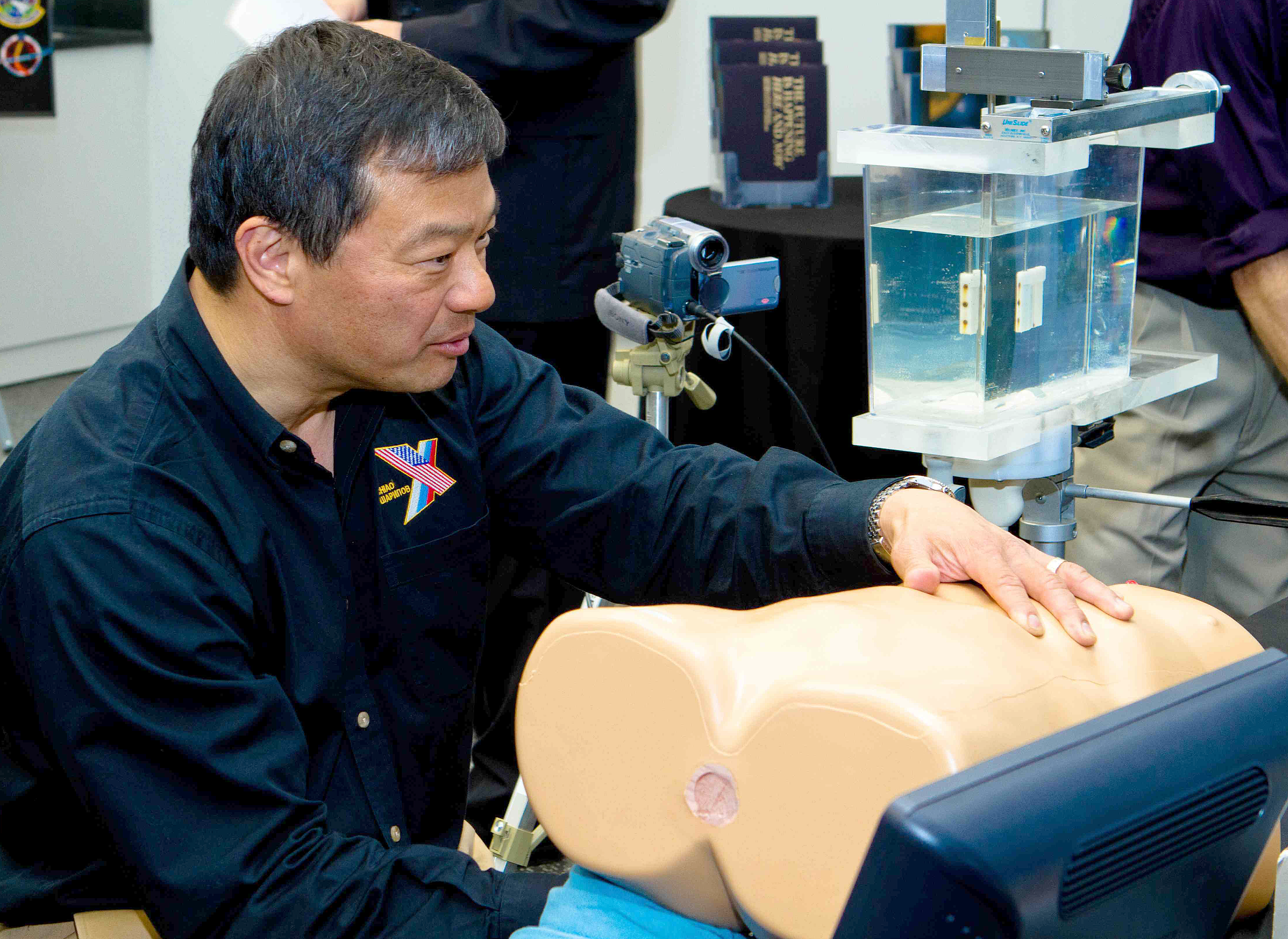
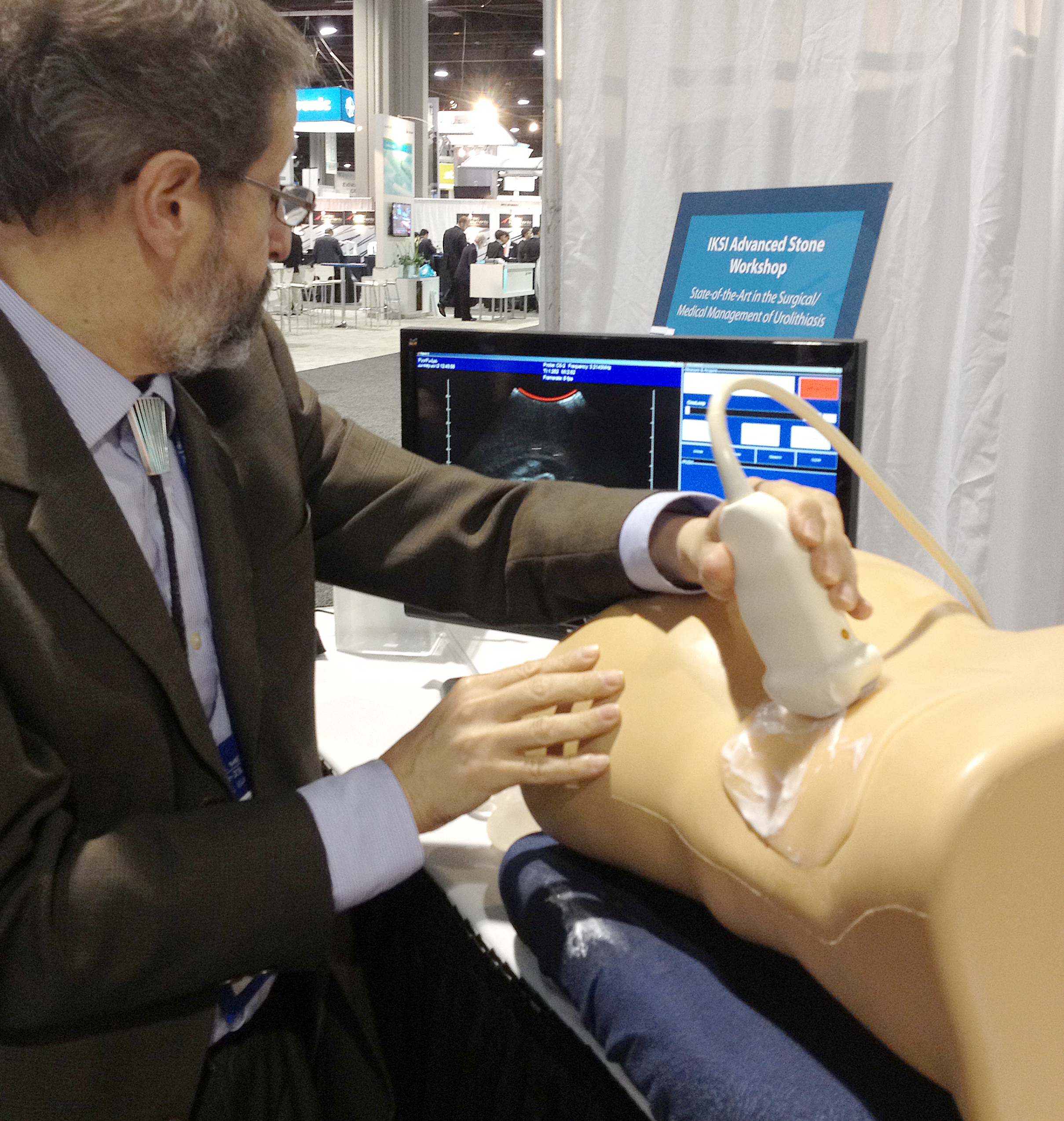
This dedicated ultrasound system is capable of non-invasive, real-time, image-guided repositioning of kidney stones. We can move stones while patients are awake so there’s no pain — this can be an office procedure. The real goal is to relocate small stones to facilitate their passage.
This is completely novel technology. There’s nothing like it out there.
|
|
Clinical Validation Study
|
|
|
Presentation by Mike Bailey, February 2021
|
|
|
Renal Stone Management Technology Development and Clinical Validation Study (PDF, 20 MB)
Presentation at Breaking Boundaries: Advancing Human Space Flight Research Through Innovation and Collaboration, organized by NASA, February 2021 |
|
 |
|
|
First-in-Human Feasibility Study
|
|
|
|
|
|
In almost every clinical trial patient, we could reposition stones with ultrasound. In one dramatic case, small stones were actually passed within hours of the study treatment.
|
|
Feasibility Study Results
|
|
|
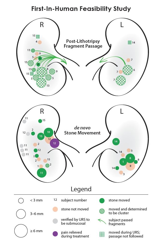
Subjects underwent ultrasonic propulsion while awake without sedation in clinic, or during ureteroscopy while anesthetized. Kidney stones were repositioned in 14 of 15 subjects. Ultrasonic propulsion was able to successfully reposition stones and facilitate the passage of fragments in humans. No adverse events were associated with the investigational procedure.
- Four of six post-lithotripsy subjects passed over 30 fragments
- Moved (nudged) stones up to 10 mm
- Relieved pain in one subject
- No safety concerns
- Diagnostic tool to distinguish a pack of small stones from a single large stone
|
First-in-human clinical trial of ultrasonic propulsion of kidney stones
Harper, J.D., et al., J. Urol. 195, 956-964, doi:10.1016/j.juro.2015.10.131, 2016. |
|
 |
|
Expelling stones with ultrasonic propulsion
R. Kelsey, Nature Rev. Urol. 13, doi:doi:10.1038/nrurol.2015.281, 2015. |
|
 |
|
Ultrasonic propulsion of kidney stones
May, P.C., M.R. Bailey, and J.D. Harper, Current Opinion in Urol. 26, 264-270, doi:10.1097/MOU.0000000000000276, 2016. |
|
 |
|
Breaking New Ground (and Kidney Stones) with Ultrasound (PDF, 3 MB)
Urology Times, November 2015, Q&A with Michael Bailey |
|
 |
|
"First-in-Humans" Clinical Trial Success
News Release from NSBRI: "First in Humans" Clinical Trial Demonstrates Non-Invasive Expulsion of Kidney Stones, 19 May 2015 |
|
 |
|
Ultrasound Able to Reposition and Facilitate Passage of Kidney Stones
MedicalResearch.com interview with Mike Bailey, 27 May 2015 |
|
 |
|
Researchers Report Results of First Clinical Trial for Ultrasonic Propulsion of Stones
AUA Daily News, May 2015 |
|
 |
|
|
|
|
RECENT POSTERS & PRESENTATIONS
|
|
|
|
WCET 2024
|
World Congress of Endourology and Uro-Technology, 12–14 August 2024, Seoul, South Korea
Ultrasound-Facilitated Clearance of Small Residual Kidney Stone Fragments Reduces Relapse Incidence
Removal of Small, Asymptomatic, Renal Stones with Burst Wave Lithotripsy and Ultrasonic Propulsion
Treating Obstructing Ureteroliths in Pet Cats with Burst Wave Lithotripsy
|
|
ACVIM Forum 2024
|
|
A Pilot Study of Burst Wave Lithotripsy for Treatment of Obstructive Ureteroliths in Cats
American College of Veterinary Internal Medicine, 6–8 June, Minneapolis |
|
 |
|
|
2024 IAAAM Conference
|
|
Application of Novel Break Wave Lithotripsy and Ultrasonic Propulsion Technology for the Treatment of Ureteral Calculi in a Bottlenose Dolphin (Tursiops truncatus) (PDF, 157 KB)
2024 International Association for Aquatic Animal Medicine Conference, 19–23 May 2024, Galway, Ireland |
|
 |
|
|
AMERICAN UROLOGICAL ASSOCIATION 2016
|
|
|
|
Download the complete and printable:
|
|
2016 PRESENTATION SCHEDULE: NIH NIDDK Program Project Group DK043881 (PDF, 841 KB)
EUS – Section of the Endourological Society, Talks and Posters & AUA Presentations |
|
 |
|
|
EUS Plenary Lectures at Marriott: Marina DE
|
|
Hands-on Demonstration Booth #4106
|
Burst Wave Lithotripsy: Noninvasive Stone Disintegration by Focused Ultrasound Without Shock Waves
Speaker: Adam Maxwell, 8:18–8:28 AM
EUS Best Abstract Award: Increased Contrast of Stone Specific Ultrasound Imaging in Human Subjects
Speaker: Philip C. May, MD, 11:30–11:50 AM
|
|
|
|
AUA Talks at SDCC
|
|
|
PD19-11: Pilot Assessment of Transcutaneous Boiling Histotripsy Ablation of the Kidney in the Porcine Model
George R. Schade et al., 1:00–3:00 PM, Room 23AB
V6-01: Introduction of a Renal Papillary Grading System for Patients with Nephrolithiasis
Michael S. Borofsky et al., 10:30 AM–12:30 PM, Room 29CD
MP51-10: Ureteral Strictures After Ureteroscopy for Nephrolithiasis: Multi-Institutional Outcomes
Philip C. May et al., 1:00–3:00 PM, Room 30ABC
MP54-02: Development of a Novel Magnetic Resonance Imaging (MRI) Acquisition and Analysis Workflow for the Quantification of Renal Hemorrhagic Injury
Paul Territo et al., 3:30–5:30 PM, Room 31
MP54-13: Detection and Assessment of Hemorrhagic Kidney Injury Caused by Burst Wave Lithotripsy Using Ultrasound and Magnetic Resonance Imaging
Adam Maxwell et al., 3:30–5:30 PM, Room 31
MP58-02: Novel Insight into Stone Formation Mechanisms Using Micro CT: Verification that Randall’s Plaques and Ductal Plugs Can Produce Clinically Significant Stones
James C. Williams, Jr. et al., 8:00–10:00 AM, Room 33
|
|
|
|
87th Annual Meeting of the Aerospace Medical Association, 24–28 April, Atlantic City, NJ
|
J.C. Simon, B.W. Cunitz, O.A. Sapozhnikov, Y.-N. Wang, F. Starr, J. Thiel, J.R. Holm, M.D. Sorensen, and M.R. Bailey. Kidney stone detection in space with the color-Doppler ultrasound twinkling artifact.
|
|
45th Annual Ultrasonic Industry Association Symposium, 4–6 April 2016, Seattle, WA
|
M. Bailey, B. Cunitz, B. Dunmire, P. May, O. Levy, H. Wessells, M. Sorensen, and J. Harper. Technology to reposition kidney stones with ultrasound.
V.A. Khokhlova, P.V. Yuldashev, P.B. Rosnitskiy, A.D. Maxwell, W. Kreider, M.R. Bailey, and O.A. Sapozhnikov. Design of HIFU transducers to generate specific nonlinear ultrasound fields.
J.C. Simon, B.W. Cunitz, J. Thiel, O.A. Sapozhnikov, J.R. Holm, B. Dunmire, M.D. Sorensen, and M.R. Bailey. Evidence for trapped bubbles as the cause for the color Doppler ultrasound twinkling artifact.
A. Maxwell, W. Kreider, Y.-N. Wang, B. Cunitz, O. Sapozhnikov, M. Hubbard, B. Dunmire, M. Bailey, J. Harper, and M. Sorensen. Burst wave lithotripsy: A noninvasive method to fragment kidney stones with sinusoidal ultrasound pulses.
|
|
American Institute of Ultrasound in Medicine, 17–21 March 2016, New York
|
|
First-in-human clinical trial using ultrasonic propulsion to reposition renal calculi in humans
J. Thiel, J.D. Harper, B.W. Cunitz, B. Dunmire, F. Lee, M.D. Sorensen, R. Hsi, H. Wessells, J.E. Lingeman, and M.R. Bailey |
|
 |
|
|
16th International Symposium on Therapeutic Ultrasound, 14–16 March 2016, Tel Aviv, Israel
|
|
Comparison of derating methods for nonlinear ultrasound fields of diagnostic-type transducers
Yldashev, P.V., M.M. Karzova, B.W. Cunitz, B. Dunmire, W. Kreider, O.A. Sapozhnikov, M.R. Bailey, and V.A. Khokhlova |
|
 |
|
|
Human Research Program Investigators' Workshop, 8–11 February 2016, Galveston, TX
|
B.W. Cunitz, A. Maxwell, B. Dunmire, B. MacConaghy, M.R. Bailey, O. Levy, J. Harper, and M. Sorensen. Prevention of renal stone complications in space exploration.
|
|
21st European Symposium on Ultrasound Contrast Imaging, 21–22 January 2016, Rotterdam, The Netherlands
|
M. Bruce, B. Cunitz, C.T. Darveau, J. Simon, B. Dunire, and M. Bailey. Observations of the Doppler twinkling artifact of kidney stones is bubble related.
|
|
9th International Symposium on Cavitation, 6–10 December 2015, Lausanne, Switzerland
|
|
Modeling and experimental analysis of acoustic cavitation bubbles for burst wave lithotripsy
K. Maeda, T. Colonius, W. Kreider, A. Maxwell, B. Cunitz, and M. Bailey |
|
 |
|
|
20th International Symposium on Nonlinear Acoustics, 29 June – 3 July 2015, Lyon, France
|
|
Nonlinear effects in ultrasound fields of diagnostic-type transducers used for kidney stone proulsion: Characterization in water
M. Karzova, B. Cunitz, P. Yuldasheve, Y. Andriyakhina, W. Kreider, O. Sapozhnikov, M. Bailey, and V. Khokhlova |
|
 |
|
|
3rd International Consultation on Stone Disease, 12–15 October, Glasgow, Scotland
|
J.E. Lingeman, et al., including M.R. Bailey. Stone technology: Shock wave and intracorporeal lithotripsy. International Consultation on Urological Diseases – Societe Internationale Urologique (Paris)
|
|
IEEE International Ultrasonics Symposium, 3–6 September 2014, Chicago, IL
|
Barbrina Dunmire, Franklin Lee, Bryan Cunitz, Jonathan Harper, Mathew Sorensen, Marla Paun, and Michael Bailey. Using Ultrasound to determine the size of kidney stones.
Michael Bailey, Franklin Lee, Bryan Cunitz, Barbrina Dunmire, Marla Paun, Susan Ross, James E. Lingeman, Michael Coburn, Hunter Wessells, Mathew Sorensen, and Jonathan Harper, Ultrasonic propulsion of kidney stones: Preliminary results from human feasibility study.
Julianna C. Simon, Oleg A. Sapozhnikov, Vera A. Khokhlova, Yak-Nam Wang, Lawrence A. Crum, and Michael R. Bailey. The role of cavitation in the ultrasonic atomization of liquids and tissues.
Oleg Sapozhnikov, Adam Maxwell, Wayne Kreider, and Michael Bailey. Solid particle transverse trapping at the focus of 1.5-MHz vortex beam radiated by 12-sector ultrasonic array.
Wayne Kreider, Adam D. Maxwell, Bryan Cunitz, Yak-Nam Wang, Ryan Hsi, Franklin Lee, Mathew Sorensen, Jonathan Harper, Vera A. Khokhlova, Bret A. Connors, Andrew P. Evan, and Michael R. Bailey. In vivo evaluation of cavitation activity and hemorrhagic kidney injury by burst wave lithotripsy.
Bryan W. Cunitz, John C. Kucewicz, Barbrina Dunmire, Marla Paun, Ryan Hsi, Franklin Lee, Jonathan D. Harper, Mathew D. Sorensen, Oleg A. Sapozhnikov, and Michael R. Bailey. Real-time kidney stone detection using optimized Doppler imaging.
Michael Bailey, Franklin Lee, Ryan Hsi, Marla Paun, Barbrina Dunmire, Ziyue Liu, Jonathan Harper, and Mathew Sorensen. Shockwave lithotripsy with renoprotective pause is associated with renovascular vasoconstriction in humans.
|
2014 IEEE IUS website
|
|
 |
|
|
International Symposium on Ultrasonic Imaging and Tissue Characterization, 9–11 June 2014, Arlington, VA
|
Bryan W. Cunitz, John C. Kucewicz, Barbrina Dunmire, Marla Paun, Franklin Lee, Jonathan D. Harper, Mathew D. Sorensen, Oleg A. Sapozhnikov, and Michael R. Bailey. Optimization of Doppler ultrasound imaging to enhance real-time kidney stone detection.
Barbrina Dunmire, Franklin Lee, Bryan Cunitz, Jonathan Harper, Mathew Sorensen, Marla Paun, and Michael Bailey. Ultrasonic sizing of kidney stones.
|
2014 UITC website
Click on 2014 abstracts and search for APL-UW authors to read complete abstracts. |
|
 |
|
|
American Urological Association Annual Meeting, 16–21 May, Orlando, FL
|
George R. Schade, Adam D. Maxwell, Yak-Nam Wang, Tatiana D. Khokhlova, Daniel W. Lin, Oleg A. Sapozhnikov, Michael R. Bailey, and Vera A. Khokhlova. Pilot evaluation of boiling histotripsy of the kidney: Assessment in human ex vivo kidneys and validation of the porcine model.
Jonathan Harper, Franklin Lee, Bryan Cunitz, Barbrina Dunmire, Marla Paun, Susan Ross, Michael Bailey, James E. Lingeman, Michael Coburn, Hunter Wessells, and Mathew Sorensen. Ultrasonic propulsion of kidney stones: Preliminary results from human feasibility study.
Adam D. Maxwell, Franklin Lee, Bryan W. Cunitz, Barbrina Dunmire, Wayne Kreider, Mathew D. Sorensen, Michael R. Bailey, and Jonathan D. Harper. In vitro fragmentation of renal calculi by burst wave lithotripsy: Effect of stone composition.
Franklin C. Lee, Bryan Cunitz, Barbrina Dunmire, Mathew Sorensen, Jonathan Harper, and Michael Bailey. Novel automated stone detection system to measure renal calculi with ultrasound.
Franklin C. Lee, Barbrina Dunmire, Jonathan Harper, Bryan Cunitz, Michael Bailey, and Mathew Sorensen. The acoustic shadow width is a more accurate predictor of true stone size during ultrasound.
Franklin Lee, Ryan Hsi, Mathew Sorensen, Marla Paun, Barbrina Dunmire, Ziyue Liu, Michael Bailey, and Jonathan Harper. Shockwave lithotripsy with renoprotective pause is associated with renovascular vasoconstriction in humans.
Adam D. Maxwell, Wayne Kreider, Bryan W. Cunitz, Yak-Nam Wang, Ryan S. Hsi, Franklin C. Lee, Mathew D. Sorensen, Jonathan D. Harper, and Michael R. Bailey. Evaluation of stone comminution and tissue injury in vivo using a novel method of extracorporeal lithotripsy without shock waves.
|
AUA Annual Meeting website
|
|
 |
|
|
Acoustical Society of America Spring Meeting, 5–9 May, Providence, RI
|
Julianna Simon, Oleg Sapozhnikov, Vera Khokhlova, Yak-Nam Wang, Wayne Kreider, Lawrence Crum, and Michael Bailey. The role of bubbles in the atomization of liquids and tissues.
Franklin C. Lee, Barbrina Dunmire, Jonathan D. Harper, Bryan W. Cunitz, Marla Paun, Michael R. Bailey, and Mathew D. Sorensen, Ultrasound acoustic shadow width is an accurate predictor of kidney stone size.
|
ASA Spring 2014 Meeting website
|
|
 |
|
|
International Society for Therapeutic Ultrasound Symposium, 2–5 April, Las Vegas, NV
|
Julianna C. Simon, Oleg A. Sapozhnikov, Vera A. Khokhlova, Yak-Nam Wang, Lawrence A. Crum, and Michael Bailey. Ultrasonic atomization: A mechanism of tissue fractionation in boiling histotripsy.
Wayne Kreider, Adam D. Maxwell, Bryan Cunitz, Yak-Nam Wang, Ryan Hsi, Franklin Lee, Mathew Sorensen, Jonathan Harper, Vera A. Khokhlova, Andrew P. Evan, and Michael R. Bailey. A preliminary assessment of the potential for kidney injury by burst wave lithotripsy.
Yak-Nam Wang, Tatiana Khokhlova, Adam Maxwell, Wayne Kreider, Ari Partanen, Navid Farr, G. Schade, Michael Bailey, and Vera Khokhlova. Mechanical decellularization of tissue while sparing vascular structures using boiling histotripsy.
Petr V. Yuldashev, Adam D. Maxwell, Wayne Kreider, Tatiana D. Khokhlova, Oleg A. Sapozhnikov, Michael R. Bailey, Lawrence A. Crum, and Vera A. Khokhlova. Acoustic field of a therapeutic transducer for generating boiling histotripsy lesions at significant depths in tissue: Combined measurement and modeling characterization.
Adam D. Maxwell, Bryan W. Cunitz, Wayne Kreider, Oleg A. Sapozhnikov, Ryan S. Hsi, Mathew D. Sorensen, Jonathan D. Harper, and Michael R. Bailey. Burst wave lithotripsy: A new method of stone fragmentation without shock waves.
Franklin Chong-Ho Lee, Yak-Nam Wang, Barbrina Dunmire, Julianna C. Simon, H. Denny Liggitt, Marla Paun, Bryan W. Cunitz, Frank Starr, Michael R. Bailey, Kristina Penniston, Ryan S. Hsi, Mathew D. Sorensen, and Jonathan D. Harper. Preclinical safety and effectiveness studies of ultrasonic propulsion of kidney stones.
|
The 14th ISTU Symposium website
|
|
 |
|
|
NASA Human Research Program Investigators' Workshop, 12–13 February 2014, Galveston, TX
|
B.L. Dunmire, J.D. Harper, M.D. Sorensen, H. Wessells. J.E. Lingeman, M. Coburn, B. W. Cunitz, Y.-N. Wang, J.C. Simon, A.D. Maxwell, W. Kreider, M. Paun, L.A. Crum, V.A. Khokhlova, O.A. Sapozhnikov, and M.R. Bailey. Prevention of renal stone complications in space exploration.
J.C. Simon, O.A. Sapozhnikov, V.A. Khokhlova, Y.-N. Wang, L.A. Crum, and M.R. Bailey. Ultrasonic atomization of tissue: A mechanism for ultrasound-based surgery.
|
NASA HRPI Workshop website
Click on Program and Abstracts and then search the Author Index |
|
 |
|
|
31st World Congress of Endourology & SWL, 22–26 October 2013, New Orleans, LA
|
Ryan S. Hsi, Barbrina Dunmire, Bryan W. Cunitz, Xuemei He, Mathew D. Sorensen, Jonathan D. Harper, Michael R. Bailey, and Thomas S. Lendvay. Content and face validation of a curriculum for ultrasonic propulsion of renal calculi in a human phantom.
Adam D. Maxwell, Bryan W. Cunitz, Wayne Kreider, Oleg A. Sapozhnikov, Ryan S. Hsi, Mathew D. Sorensen, Jonathan D. Harper, and Michael R. Bailey. Novel method of extracorporeal lithotripsy without shock waves: In vitro fragmentation of artificial and human calculi.
Adam D. Maxwell, Ryan S. Hsi, Michael R. Bailey, Pasquale Casale, and Thomas S. Lendvay. Cavitation-based focused ultrasound for noninvasive puncture of ureteroceles: In vitro results.
Ryan S. Hsi, Bryan W. Cunitz, Barbrina Dunmire, Marla Paun, Jonathan D. Harper, Michael R. Bailey, and Mathew D. Sorensen. Development of dedicated stone detection protocols using a research-based ultrasound imager.
|
WCE & SWL Congress website and official program
|
|
 |
|
|
166th Meeting of the Acoustical Society of America, 2–6 December 2013, San Francisco, CA
|
 |
|
Addressing nonlinear propagation effects in characterization of high intensity focused ultrasound fields and prediction of thermal and mechanical bioeffects in tissue Khokhlova, V.A., P.V. Yuldashev, W. Kreider, O.A. Sapozhnikov, M.R. Bailey, T.D. Khokhlova, A.D. Maxwell, and L.A. Crum, "Addressing nonlinear propagation effects in characterization of high intensity focused ultrasound fields and prediction of thermal and mechanical bioeffects in tissue," J. Acoust. Soc. Am., 134, 4153, doi:10.1121/1.4831221, 2013. |
|
More Info
| |
|
1 Nov 2013 
|
|
 |
|
|
Nonlinear propagation effects are present in most fields generated by high intensity focused ultrasound (HIFU) sources. In some newer HIFU applications, these effects are strong enough to result in the formation of high amplitude shocks that actually determine the therapy and provide a means for imaging. However, there is no standard approach yet accepted to address these effects. Here, a set of combined measurement and modeling methods to characterize nonlinear HIFU fields in water and predict acoustic pressures in tissue is presented. A characterization method includes linear acoustic holography measurements to set a boundary condition to the model and nonlinear acoustic simulations in water for increasing pressure levels at the source. A derating method to determine nonlinear focal fields with shocks in situ is based on the scaling of the source pressure for data obtained in water to compensate for attenuation losses in tissue. The accuracy of the methods is verified by comparing the results with hydrophone and time-to-boil measurements. Major effects associated with the formation of shocks are overviewed. A set of metrics for determining thermal and mechanical bioeffects is introduced and application of the proposed tools to strongly nonlinear HIFU applications is discussed.
|
|
 |
|
An ultrasound system to identify and characterize kidney stones Cunitz, B.W., B.L. Dunmire, M.D. Sorensen, R. Hsi, F. Lee, O.A. Sapozhnikov, J.D. Harper, and M. Bailey, "An ultrasound system to identify and characterize kidney stones," J. Acoust. Soc. Am., 134, 3976, doi:10.1121/1.4830485, 2013. |
|
More Info
| |
|
1 Nov 2013 
|
|
 |
|
|
Ultrasound imaging has tissue and blood imaging modes. This report describes development of a kidney stone imaging mode. Two plane pulses generate a B-mode image. Overlaid in color are regions of high decorrelation between the pulses. Our previous data [UMB, 39, 1026-1038 (2013)] indicate the pulses excite bubbles on the stone surface, which causes the decorrelation. As such this mode automatically identifies stones in the image while scanning at a high frame rate. Further in a control box placed on the stone, highly focused beams are scanned across the stone and a harmonic B-mode image is produced to sharpen the lateral resolution. This mode is used to refine the size and shape of the stone. The first mode is used to aid visualization of stones. Our team is also using it to target and track stones that move with respiration during shock wave lithotripsy (SWL) and as an indicator of stone susceptibility to SWL since surface bubbles contribute to comminution. Improved stone sizing by the second mode aids treatment planning, and resolution of surface roughness is another indicator of stone fragility.
|
|
 |
|
Fragmentation of kidney stones in vitro by focused ultrasound bursts without shock waves Maxwell, A.D., B.W. Cunitz, W. Kreider, O.A. Sapozhnikov, R.S. Hsi, M.D. Sorensen, J.D. Harper, and M.R. Bailey, "Fragmentation of kidney stones in vitro by focused ultrasound bursts without shock waves," J. Acoust. Soc. Am., 134, 4183, doi:10.1121/1.4831340, 2013. |
|
More Info
| |
|
1 Nov 2013 
|
|
 |
|
|
Shock wave lithotripsy (SWL) is the most common procedure for treatment of kidney stones. SWL noninvasively delivers high-energy focused shocks to fracture stones into passable fragments. We have recently observed that lower-amplitude, sinusoidal bursts of ultrasound can generate similar fracture of stones. This work investigated the characteristics of stone fragmentation for natural (uric acid, struvite, calcium oxalate, and cystine) and artificial stones treated by ultrasound bursts. Stones were fixed in position in a degassed water tank and exposed to 10-cycle bursts from a 200-kHz transducer with a pressure amplitude of p ≤ 6.5 MPa, delivered at a rate of 40–200 Hz. Exposures caused progressive fractures in the stone surface leading to fragments up to 3 mm. Treatment of artificial stones at different frequencies exhibited an inverse relationship between the resulting fragment sizes and ultrasound frequency. All artificial and natural types of stones tested could be fragmented, but the comminution rate varied significantly with stone composition over a range of 12–630 mg/min. These data suggest that stones can be controllably fragmented by sinusoidal ultrasound bursts, which may offer an alternative treatment strategy to SWL.
|
|
 |
|
Holography and numerical projection methods for characterizing the three-dimensional acoustic fields of arrays in continuous-wave and transient regimes Kreider, W., A.D. Maxwell, P.V. Yuldashev, B.W. Cunitz, B. Dunmire, O.A. Sapozhnikov, and V.A. Khokhlova, "Holography and numerical projection methods for characterizing the three-dimensional acoustic fields of arrays in continuous-wave and transient regimes," J. Acoust. Soc. Am., 134, 4153, doi:10.1121/1.4831222, 2013. |
|
More Info
| |
|
1 Nov 2013 
|
|
 |
|
|
The use of projection methods is increasingly accepted as a standard way of characterizing the 3D fields generated by medical ultrasound sources. When combined with hydrophone measurements of pressure amplitude and phase over a surface transverse to the wave propagation, numerical projection can be used to reconstruct 3D fields that account for operational details and imperfections of the source. Here, we use holography measurements to characterize the fields generated by two array transducers with different geometries and modes of operation. First, a seven-element, high-power therapy transducer is characterized in the continuous-wave regime using holography measurements and nonlinear forward-projection calculations. Second, a C5-2 imaging probe (Philips Healthcare) with 128 elements is characterized in the transient regime using holography measurements and linear projection calculations. Results from the numerical projections for both sources are compared with independent hydrophone measurements of select waveforms, including shocked focal waveforms for the therapy transducer. Accurate 3D field representations have been confirmed, though a notable sensitivity to hydrophone calibrations is revealed. Uncertainties associated with this approach are discussed toward the development of holography measurements combined with numerical projections as a standard metrological tool.
|
|
 |
|
Kidney stone fracture by surface waves generated with focused ultrasound tone bursts Sapozhnikov, O.A., A.D. Maxwell, W. Kreider, B.W. Cunitz, and M.R. Bailey, "Kidney stone fracture by surface waves generated with focused ultrasound tone bursts," J. Acoust. Soc. Am., 134, 4184, doi:10.1121/1.4831341, 2013. |
|
More Info
| |
|
1 Nov 2013 
|
|
 |
|
|
Previous studies have provided insight into the physical mechanisms of stone fracture in shock wave lithotripsy. Broadly focused shocks efficiently generate shear waves in the stone leading to internal tensile stresses, which in concert with cavitation at the stone surface, cause cracks to form and propagate. Here, we propose a separate mechanism by which stones may fragment from sinusoidal ultrasound bursts without shocks. A numerical elastic wave model was used to simulate propagation of tone bursts through a cylindrical stone at a frequency between 0.15 and 2 MHz. Results suggest that bursts undergo mode conversion into surface waves on the stone that continually create significant stresses well after the exposure is terminated. Experimental exposures of artificial cylindrical stones to focused burst waves in vitro produced periodic fractures along the stone surface. The fracture spacing and resulting fragment sizes corresponded well with the spacing of stresses caused by surface waves in simulation at different frequencies. These results indicate surface waves may be an important factor in fragmentation of stones by focused tone bursts and suggest that the resulting stone fragment sizes may be controlled by ultrasound frequency.
|
|
|
|
More
|
Vera A. Khokhlova, Yak-Nam Wang, Sergey V. Buravkov, Adam D. Maxwell, Tatiana D. Khokhlova, Daniel W. Lin, Oleg A. Sapozhnikov, Michael R. Bailey, and George R. Schade. Histological analysis of mechanically fractionation lesions generated in ex-vivo human and porcine kidneys using high intensity focused ultrasound. Russian Acoustical Conference, Moscow, October 2014.
Michael Bailey, Bryan Cunitz, Barbrina Dunmire, Yak-Nam Wang, Adam Maxwell, Oleg Sapozhnikov, Jonathan Harper, Mathew Sorensen, Ryan Hsi, Franklin Lee, and Lawrence Crum. Stone specific ultrasound mode for the diagnoses and treatment of kidney stones in space. 85th Annual Scientific Meeting of the Aerospace Medical Association, 11–15 May 2014, San Diego, CA.
Franklin Lee, Jonathan Harper, Ryan Hsi, Marla Paun, Barbrina Dunmire, Bryan Cunitz, Michael Bailey, and Mathew Sorensen. Stone size measurement using the ultrasound acoustic shadow. Northwest Urological Society Conference 6–7 December 2013, Vancouver, BC.
Franklin Lee, Ryan Hsi, Mathew Sorensen, Marla Paun, Barbrina Dunmire, Ziyue Liu, Michael Bailey, and Jonathan Harper. Shock wave lithotripsy pause results in vasoconstriction and theoretical renal protection in humans. Northwest Urological Society Conference 6–7 December 2013, Vancouver, BC.
Franklin Lee, Mathew D. Sorensen, Ryan S. Hsi, Barbrina Dunmire, Bryan W. Cunitz, Marla Paun, Michael R. Bailey, and Jonathan D. Harper. Development of automated kidney stone detection using a research-based ultrasound imager. Western Section American Urological Association Annual Meeting, 3–7 November 2013, Monterey, CA.
Adam D. Maxwell, Bryan W. Cunitz, Wayne Kreider, Oleg A. Sapozhnikov, Ryan S. Hsi, Mathew D. Sorensen, Jonathan D. Harper, and Michael R. Bailey. Extracorporeal lithotripsy using ultrasound pulses without shock waves: In vitro results. Western Section American Urological Association Annual Meeting, 3–7 November 2013, Monterey, CA.
|
|
Patents Issued + Invention Disclosures to UW CoMotion
|
 |
|
Noninvasive Fragmentation of Urinary Tract Stones with Focused Ultrasound Patent Number: 12,167,864 |
|
More Info
| |
|
17 Dec 2024 
|
|
 |
|
|
A method for attempting to fragment or comminute an object in a body using ultrasound includes producing a burst wave lithotripsy (BWL) waveform by a therapy transducer. The BWL waveform is configured to fragment or comminute the object. The BWL waveform includes a first burst of continuous ultrasound cycles and a second burst of continuous ultrasound cycles. A burst frequency corresponds to a frequency of repeating the bursts of the BWL waveform. The method also includes determining a cycle frequency f of the continuous ultrasound cycles within the first burst and the second burst based on a target fragment size D, where the cycle frequency is: f(MHz)=0.47/D(mm).
|
|
 |
|
Noninvasive Fragmentation of Urinary Tract Stones with Focused Ultrasound Patent Number: 11,583,299 |
|
21 Feb 2023 
|
|
 |
 |
|
Ultrasound Based Method and Apparatus for Stone Detection and to Facilitate Clearance Thereof Patent Number: 10,039,562 |
|
More Info
| |
|
7 Aug 2018 
|
|
 |
|
|
Described herein are methods and apparatus for detecting stones by ultrasound, in which the ultrasound reflections from a stone are preferentially selected and accentuated relative to the ultrasound reflections from blood or tissue. Also described herein are methods and apparatus for applying pushing ultrasound to in vivo stones or other objects, to facilitate the removal of such in vivo objects.
|
|
 |
|
Holographic Beam Shaping for Ultrasound Therapy Transducers Record of Invention Number: 48221 |
|
1 Dec 2017 
|
|
 |
 |
|
Ultrasound Based Method and Apparatus for Stone Detection and to Facilitate Clearance Thereof Patent Number: 9,597,103 |
|
More Info
| |
|
21 Mar 2017 
|
|
 |
|
|
Described herein are methods and apparatus for detecting stones by ultrasound, in which the ultrasound reflections from a stone are preferentially selected and accentuated relative to the ultrasound reflections from blood or tissue. Also described herein are methods and apparatus for applying pushing ultrasound to in vivo stones or other objects, to facilitate the removal of such in vivo objects.
|
|
 |
|
Supplemental Know How for Pushing, Imaging, and Breaking Kidney Stones Record of Invention Number: 47878 |
|
9 Nov 2016 
|
|
 |
 |
|
Combination Burst Wave Lithotripsy and Ultrasonic Propulsion for Improved Stone Fragmentation Record of Invention Number: 47817 |
|
9 Sep 2016 
|
|
 |
 |
|
Improved Detection of Kidney Stones with Ultrasound Record of Invention Number: 47629 |
|
19 Feb 2016 
|
|
 |
 |
|
Ultrasound based method and apparatus for stone detection and to facilitate clearance thereof Patent Number: 9,204,859 |
|
More Info
| |
|
8 Dec 2015 
|
|
 |
|
|
Described herein are methods and apparatus for detecting stones by ultrasound, in which the ultrasound reflections from a stone are preferentially selected and accentuated relative to the ultrasound reflections from blood or tissue. Also described herein are methods and apparatus for applying pushing ultrasound to in vivo stones or other objects, to facilitate the removal of such in vivo objects.
|
|
 |
|
Novel Probe and Workflow for Ultrasonic Propulsion Record of Invention Number: 47322 |
|
1 May 2015 
|
|
 |
 |
|
Methods to Determine Optimal Ultrasound Pulse Parameters to Fragment Urinary Calculi Using Acoustic Feedback Record of Invention Number: 47078 |
|
6 Oct 2014 
|
|
 |
 |
|
Applications of Ultrasonic Propulsion Record of Invention Number: 47073 |
|
3 Oct 2014 
|
|
 |
 |
|
Assortment of Push Profiles for Pushing a Variety of Kidney Stones Record of Invention Number: 47072 |
|
3 Oct 2014 
|
|
 |
 |
|
Single Element Broadly Focused Ultrasonic Propulsion Device Record of Invention Number: 47074 |
|
3 Oct 2014 
|
|
 |
 |
|
Ultrasound to rotate an obstructing kidney stone Record of Invention Number: 47066 |
|
29 Sep 2014 
|
|
 |
 |
|
Ultrasound to Detach Kidney Stones from Plaque or Tissue Record of Invention Number: 46981 |
|
13 Jun 2014 
|
|
 |
 |
|
Ultrasound Instrumentation for Ureteroscopic and Transcutaneous Kidney Stone Removal Record of Invention Number: 46839 |
|
4 Feb 2014 
|
|
 |
 |
|
Ultrasound Based Method and Apparatus to Determine the Size of Kidney Stone Fragments Before Removal via Ureteroscopy Patent Number: 8,607,634 |
|
More Info
| |
|
17 Dec 2013 
|
|
 |
|
|
A transducer is used to send an ultrasound pulse toward a stone and to receive ultrasound reflections from the stone. The recorded time between a pulse that is reflected from the proximal surface and a pulse that is reflected either from the distal surface of the stone or from a surface supporting the stone is used to calculate the stone size. The size of the stone is a function of the time between the two pulses and the speed of sound through the stone (or through the surrounding fluid if the second pulse was reflected by the surface supporting the stone). This technique is equally applicable to measure the size of other in vivo objects, including soft tissue masses, cysts, uterine fibroids, tumors, and polyps.
|
|
 |
|
Method and Apparatus to Detect the Fragmentation of Kidney Stones by Measuring Acoustic Scatter Patent Number: 8,535,250 |
|
More Info
| |
|
17 Sep 2013 
|
|
 |
|
|
During shock wave therapy, a determination is made that a kidney stone has begun to fracture, and then a progress of its fragmentation is assessed. This determination can reduce the number of shock waves used to disintegrate kidney stones, and thereby reduce dose-dependent tissue damage. The identification of fracture is possible through the detection and analysis of resonant acoustic scattering, which is the radiation caused by reverberations within a stone particle that is struck by a shock wave. The scattering frequency can provide both an indication that the kidney stone has fragmented, and an indication of the relative sizes of the fragments. Such techniques can be combined with vibro-acoustography based gating that better targets the stone.
|
|
 |
|
Device and Procedure for Noninvasive Removal of Ureteral Stents Record of Invention Number: 46501 |
|
9 May 2013 
|
|
 |
 |
|
Noninvasive Fragmentation of Urinary Tract Stones with Focused Ultrasound Record of Invention Number: 46460 |
|
28 Mar 2013 
|
|
 |
 |
|
Method for Testing Output Force of an Ultrasonic Propulsion Device Record of Invention Number: 46441 |
|
12 Mar 2013 
|
|
 |
 |
|
Noninvasive Treatment of Ureteroceles with Focused Ultrasound Record of Invention Number: 46404 |
|
14 Feb 2013 
|
|
 |
 |
|
Method of Detecting Microbubbles in Tissue and Tissue Phantoms Using "Twinkling" Artifact of Doppler Imaging Record of Invention Number: 46179 |
|
10 Aug 2012 
|
|
 |
 |
|
Using Bubbles to Better Detect Kidney Stones Record of Invention Number: 46062 |
|
30 Apr 2012 
|
|
 |
 |
|
Additional Details for Ultrasonic Propulsion Record of Invention Number: 46012 |
|
21 Mar 2012 
|
|
 |
 |
|
New Probe for Ultrasonic Propulsion of Kidney Stones Record of Invention Number: 46006 |
|
20 Mar 2012 
|
|
 |
 |
|
An Ultrasound Phantom for Detecting and Repositioning Kidney Stones Record of Invention Number: 45981 |
|
1 Mar 2012 
|
|
 |
 |
|
A New Ultrasound Imaging Regime for Improved Size Measuring of Hard Concretions Present in Soft Tissue Based on Observation of Ultrasound Shadow on a B-mode Image Record of Invention Number: 45653 |
|
8 Jun 2011 
|
|
 |
 |
|
New Regime of Ultrasound Imaging of Strong Scatterers in Tissue Using Envelope-based Beamforming Record of Invention Number: 45654 |
|
8 Jun 2011 
|
|
 |
 |
|
Ultrasound Technique to Separate Hard Objects from Tissue by Long Lasting Reverberation in Hard Objects Record of Invention Number: 45655 |
|
8 Jun 2011 
|
|
 |
 |
|
Method for Kidney Stone Detection and Targeting Using Amplitude Mode (A-mode) Ultrasound Imaging with Application to Shock Wave Lithotripsy Record of Invention Number: 8683D |
|
11 May 2010 
|
|
 |
 |
|
Method of Detecting Kidney Stones Using Ultrasound Record of Invention Number: 8633D |
|
26 Mar 2010 
|
|
 |
 |
|
Improved Detection of Hard Concretions Present in Soft Tissues Based on Doppler Imaging Twinkling Artifact by Means of Insonifying the Imaged Region with Additional Modulated Intense Ultrasound Beam Record of Invention Number: 8335D |
|
1 Apr 2009 
|
|
 |
 |
|
Improved Detection of Hard Concretions Present in Soft Tissues Based on Ultrasound Imaging Twinkling Artifact by Means of Introducing Fluctuations in Beam Structure for Consecutive Ultrasound Pulses of Each Scan Line Record of Invention Number: 8336D |
|
1 Apr 2009 
|
|
 |
|
|


